
The OCT & Retinal Camera combo is a very powerful tool. From taking standard pictures of the front and the back of the eye to scanning the 10 layers of the retina.
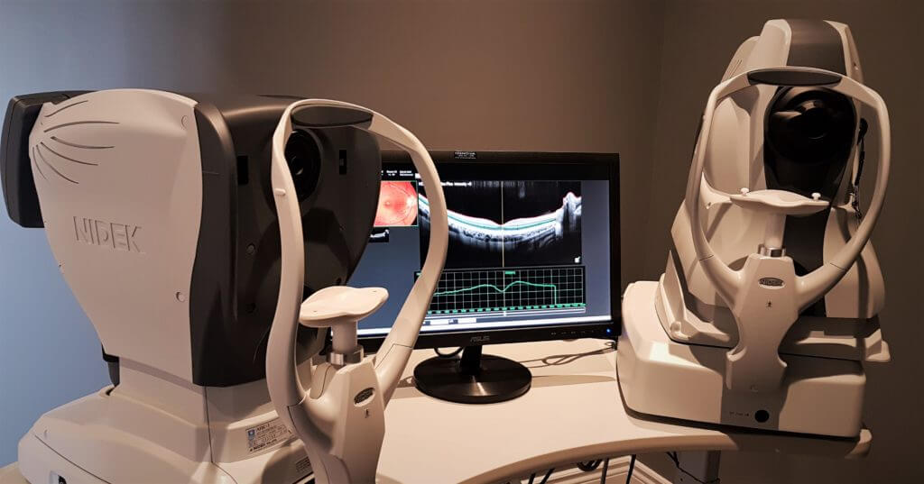
Fundus Camera
The Fundus Camera takes a picture of the back of the eye, the retina. With this image, we can see some key parts: the optic nerve, the macula and blood vessels.
With time, all three of these important ocular parts can change. For example, your optic nerve can be affected by glaucoma, your macula by macular degeneration and your blood vessels by high blood pressure or cholesterol. This isn’t even close to a full list of conditions that we can see when we examine the eyes!
Is this for you?
We recommend a baseline Retinal (Fundus) picture for our patients:
- 5 years old and above
- repeated at least every 2 years
OCT (posterior segment)
Similar to how an ultrasound uses ultrasound waves as a non-invasive way to create an image. The OCT uses light waves to create a visual representation of the different layers of the retina.
The OCT’s non-invasive scan of the 10 layers of the retina is a very key tool in monitoring for conditions such as: glaucoma, macular degeneration, diabetic retinopathy, central serous retinopathy and more.
Did you know? Both glaucoma and macular degeneration can lead to blindness. Also, the risk of developing them increases with age. This is one of the reasons why we offer this great technology.
Is this for you?
We recommend a baseline OCT scan for our patients:
- Adult patients
- all patients diagnosed with diabeties, glaucoma, macular degeneration or other retinal disease
- (frequency of scans will vary by patient)